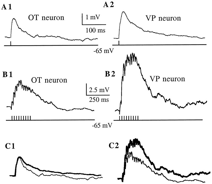
Fig. 7.
Summation of AMPA-mediated EPSPs during high-frequency synaptic stimulation. EPSPs were evoked by a single- or high-frequency (50 Hz, 200 msec) extracellular stimulation dorsolateral to the SON. Neurons were current-clamped at −70 mV. All traces were taken in the presence of ±APV (100 μm).A, Averaged EPSP (n = 60) obtained in an OT (A1) and VP (A2) neuron in response to a single shock stimulus. B, During a 50 Hz stimulation, EPSPs summed to form a slow depolarization that outlasted the duration of the stimulus. Note that the slow depolarization was larger in the VP (B2) than in the OT (B1) neuron. All traces are averages (n = 6).C, The EPSP in the VP neuron (thick line) was normalized to the amplitude of the EPSP in the OT neuron, and the traces are shown superimposed (C1). The same scaling factor was then used to scale down the envelope of the VP neuron (thick line), still showing a larger amplitude than the envelope in the OT neuron (C2).

