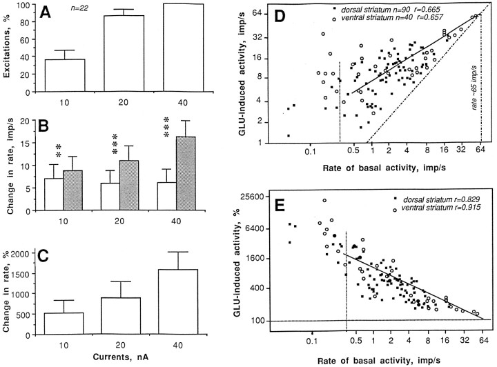
Fig. 7.
Glutamate responses of striatal neurons in awake, unrestrained rats under control conditions. Parameters of the GLU response (A, number of units with excitation, percentage; B, discharge rate before and during GLU application, impulses per second; C, relative magnitude of GLU-induced excitation, percentage) evaluated in 22 units at different GLU ejection currents (in nanoamperes). Relationships between basal discharge rate (impulses per second) and absolute (impulses per second; D) or relative changes (percentage;E) induced by GLU shown separately for units recorded from dorsal (caudoputamen) and ventral striatum (accumbens). Regression lines (common for all striatal units) are solid, and lines of no effect are hatched. n, Number of tests; r, coefficients of correlation.Vertical hatched line indicates the border between spontaneously active and sporadically active units.

