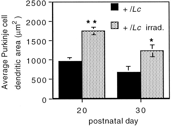
Fig. 3.
Increased Purkinje cell dendritic area in Lurcher (+/Lc) mice after the destruction of granule cells with x rays. The area encompassed by the Purkinje cell dendritic tree was measured from midsagittal cerebellar sections of P20 mice processed for anti-Calbindin immunocytochemistry. Ten to 20 Purkinje cells were analyzed per animal. Error bars indicate SEM (n = 3). Asterisks denote statistically significant differences: *p < 0.05; **p < 0.01 (unpaired two-tailed Student’s t test).

