Fig. 5.

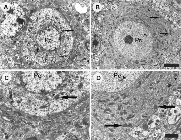
Decreased ultrastructural abnormalities in +/Lc Purkinje cells after the destruction of granule cells with x rays. The micrographs are taken from sagittal sections of the cerebellar vermis of P20 mice. Low- and high-power micrographs of a Purkinje cell in nonirradiated (A, C) and x-irradiated (B, D) Lurcher (+/Lc) mice. The Purkinje cell (Pc) in the micrographs taken from the nonirradiated +/Lc mouse displays abnormalities characteristic of the mutation: the nuclear chromatin is clumped, the nuclear membrane is irregular, and the cytoplasmic organelles are disorganized: the rough endoplasmic reticulum is not arranged into Nissl bodies, and the mitochondria are distended with spherical profiles. In contrast, the Purkinje cell (Pc) in the micrograph taken from the x-irradiated +/Lc mouse has a relatively normal appearance: the soma contains a spherical, electron-lucent nucleus (with a prominent nucleolus) that is encircled by normal-appearing mitochondria and Nissl bodies. The arrows point to swollen mitochondrial profiles. Scale bars: A, B, 4 μm;C, D, 2 μm.
