
Fig. 5.
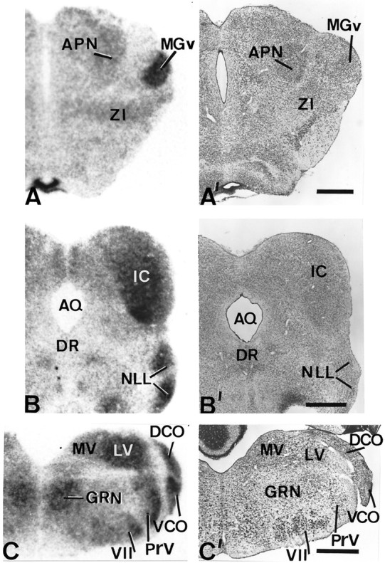
Patterns of D2 expression in the auditory pathway of hypothyroid rats. The left sideshows autoradiographs obtained from coronal sections of hypothyroid brains at thalamic (A), mesencephalic (B), and medullar (C) levels subjected to in situ hybridization. Their corresponding adjacent sections stained with cresyl violet are shown on the right side (A′, B′,C′). Note the high levels of expression in relay nuclei of the auditory pathway, i.e., the medial geniculate nucleus, the inferior colliculus, the nucleus of the lateral lemniscus, and the dorsal and ventral cochlear nuclei. DR, Dorsal nucleus raphe; MGv, medial geniculate nucleus ventral part;MV, medial vestibular nucleus; ZI, zona incerta. Scale bars, 500 μm.