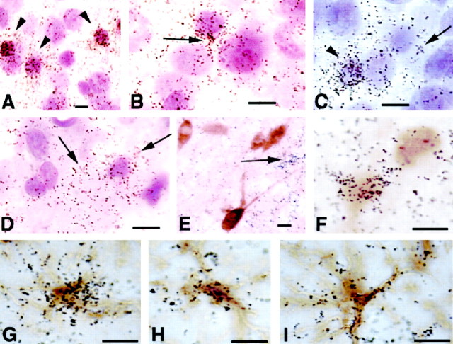
Fig. 6.
High-magnification photomicrographs taken from hypothyroid brain sections after in situhybridization. The sections were either counterstained with cresyl violet (A–D) or subjected to immunohistochemistry for calbindin (E, F) or GFAP (G–I). A, Barrel field of the somatosensory cortex; B, upper layer IV;C, deep layer IV. Note that silver grains are concentrated within small neuronal profiles (A,C, arrowheads) and in the neuropil that surrounds larger neuronal profiles, resembling glial shapes (B, C, arrows).D, Photomicrograph taken from the core of ventral posterior medial nucleus. The concentration of silver grains is mainly in the neuropil that surrounds Nissl-stained nuclei. Thearrows point to glial-like shapes. E, Several calbindin-positive, D2-negative cells photographed from layer II; D2 transcripts are distributed in the neuropil or in clusters resembling glial shapes (arrow). F, Two calbindin-positive cells photographed from layer IV of the barrel field, one of which is also positive for D2.G–I, Astrocytes from layer IV of the somatosensory cortex double-labeled for D2 byin situ hybridization and GFAP by immunohistochemistry. The silver grains are associated with the cell bodies and with the cell processes. Scale bars: A–F, 10 μm; G–I, 5 μm.

