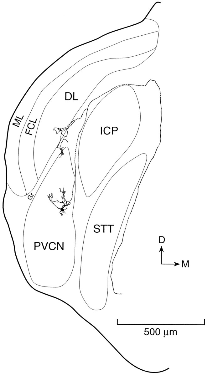
Fig. 1.

Morphology of an octopus cell labeled by intracellular perfusion with biocytin and reconstructed with a camera lucida from a series of sections cut in the same coronal plane as the original slice. A labeled octopus cell is located in the most caudal and dorsal part of the posteroventral cochlear nucleus (PVCN). The recording was made from the cell body near the caudal surface of the slice. The dendrites of the cell extend anteriorly through the thickness of the slice. Its axon exits through a fiber tract that lies at the medial surface of the dorsal cochlear nucleus and goes around the inferior cerebellar peduncle (ICP) and spinal trigeminal tract (STT). A collateral innervates granule cell regions (Gr) that lie adjacent to the octopus cell area. The molecular layer (ML), fusiform cell layer (FCL), and deep layer (DL) of the dorsal cochlear nucleus overlie the PVCN.
