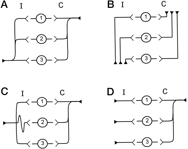
Fig. 1.
Theoretical wiring diagrams for the medial superior olive. In each panel, cells in location 1 are rostral, and cells in location 3 are caudal. Innervating axons come from either the ipsilateral (I) or contralateral (C) side. A, The wiring pattern proposed by Jeffress (1948). The ipsilateral and contralateral axons both innervate the entire length of MSO by giving rise to successive collateral branches. However, the ipsilateral axon has shorter branches caudally, whereas the contralateral axon has shorter branches rostrally. B, The Goldberg scheme (Goldberg and Brown, 1969) has different axons that innervate different rostrocaudal locations within the MSO. Different axons have different lengths.C, The avian model in which contralateral axons follow the Jeffress scheme, but the ipsilateral axon innervates the full extent of MSO with collaterals of equal length. D, A fourth wiring scheme that is a combination of the previous three models. Here, individual axons of equal length innervate the MSO from the ipsilateral side. The shortest collateral branch of a single contralateral axon innervates the rostral end of MSO before successive branches innervate central and caudal MSO. All of these schemes can create a topographical organization of ITD along the rostrocaudal dimension of MSO.

