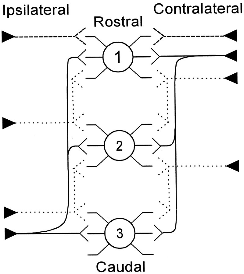
Fig. 12.
Schematic summary of the innervation patterns of the reconstructed axons from low-frequency rostral AVCN to the MSO. In a pattern similar to the Jeffress model (solid line), on the contralateral side the shortest branch terminated in the rostral end of MSO (1). Successively longer branches went to the middle (2) and caudal (3) ends of MSO. On the ipsilateral side, the shortest branch went to the caudal end of MSO with successively longer branches to central and rostral MSO, respectively. A second pattern was evident in axons with more limited termination pattern (short dashed line). Still, a third pattern was evident where individual axons terminated in restricted field (long dashed lines).

