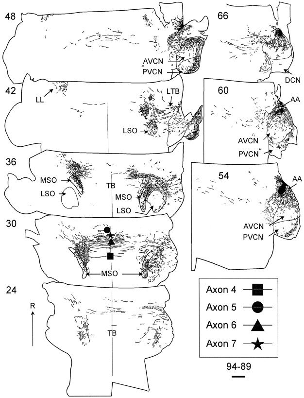
Fig. 3.
Low-magnification drawings of case 94–89 that show the location of the dextran-filled axons in the 100-μm-thick horizontal sections. Here, the more dorsal sections contain the most rostral parts of MSO. A few axons crossed the midline near the center of the rostrocaudal extent of the trapezoid body (TB), whereas none crossed caudally. Symbols in section30 show four reconstructed axons where they cross the midline (filled square, axon 4 in Fig.4C; filled circle, axon 5 in Fig.6B; filled triangle, axon 6 in Fig. 5B; filled star, axon 7 in Fig.5A). Scale bar, 1 mm. AA, Anterior part of AVCN.

