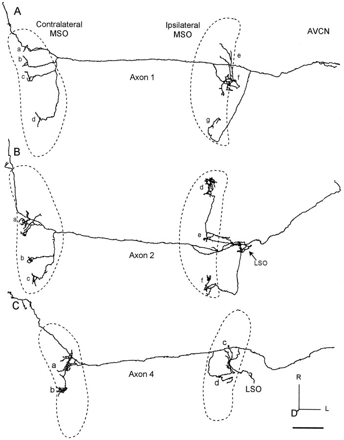
Fig. 4.
Three reconstructed axons with ladder-like branching pattern on the contralateral side and extensive branching on the ipsilateral side. The location of each MSO is indicated by thedashed line. A, Axon 1 terminated over the entire rostrocaudal length in the contralateral MSO (clusters of terminals a–d) and most of the ipsilateral side (clusters of terminalse–g). B, Axon 2 went to the central (a) and caudal parts (b, c) of contralateral MSO. Ipsilateral branches terminated in LSO (arrow) and MSO (d–f). C, Axon 4 terminated in the middle MSO on the contralateral side (a, b) and a similar area on the ipsilateral side (c, d). Scale bar, 1 mm.D, Dorsal; L, lateral.

