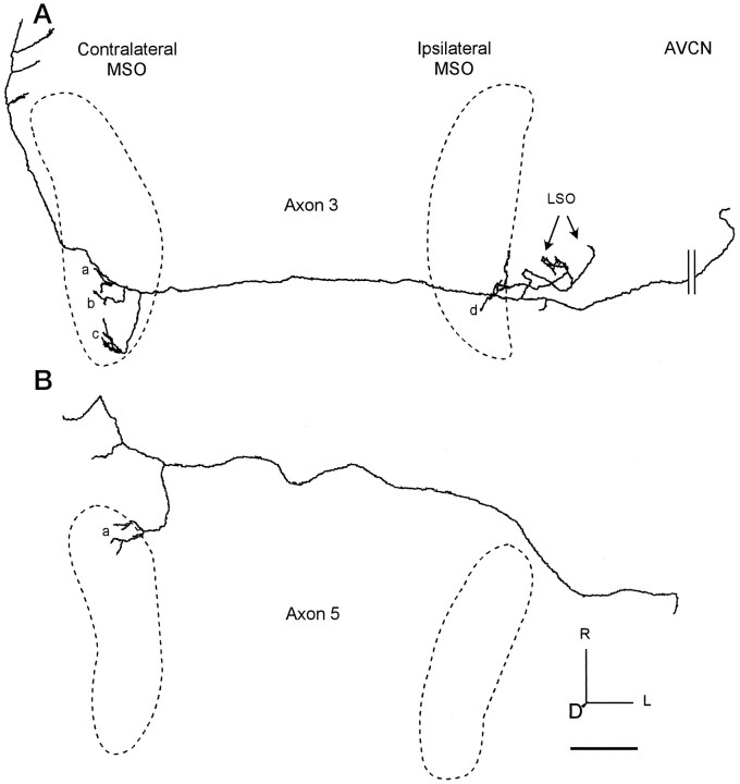
Fig. 6.
Axons with restricted branching patterns. The location of each MSO is indicated by the dashed line.A, Axon 3 terminated in caudal MSO contralaterally (clusters of terminals a–c) and ipsilaterally (e). It also terminated in LSO (arrows). B, Axon 5 terminated only in the contralateral, rostral MSO (a). The axon continued to the ventral nucleus of the lateral lemniscus, where it gave off collaterals. Scale bar, 1 mm.

