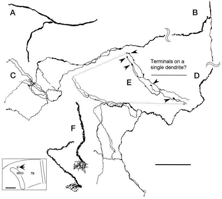
Fig. 7.
Camera lucida drawings of axon 2 in the contralateral MSO. A, First branch on contralateral side after crossing the midline trapezoid body. B, Second branch on contralateral side. C, Details of the middle terminals (Fig. 4B,b).D, Details of caudal terminals; see c in Figure 4B. E, The enlarged area ofD shows the location of five terminal boutons (arrowheads). These boutons could terminate on a single dendrite. F, Two calyceal endings in the medial trapezoid body in the superior olive contralateral to the cochlear nucleus injection. Both endings were seen in the same section of case 94–89. Both pieces were well filled at their terminals but could not be traced over any appreciable distance. Insert shows the position of calyceal endings (arrow) located medial to the rostral end of MSO. Scale bars:A–D, 100 μm; E, 50 μm; insert, 1 mm.

