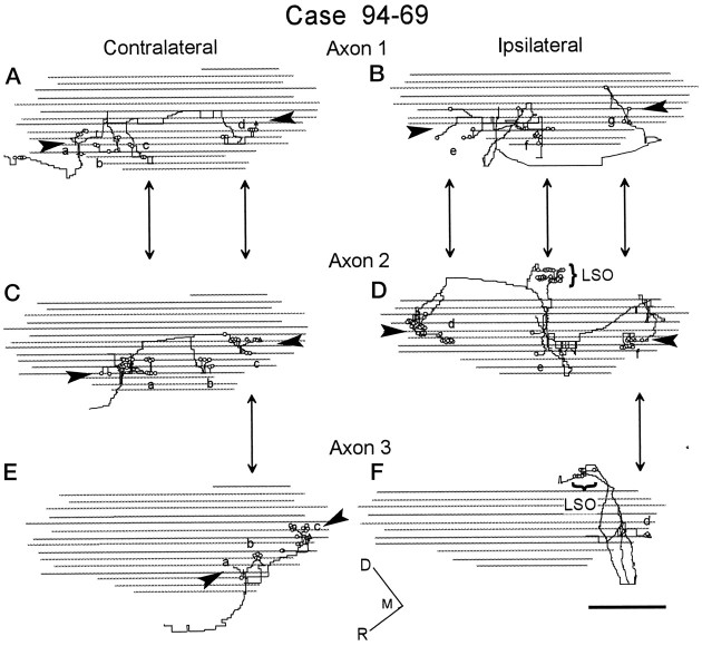
Fig. 9.
Sagittal views of each half of the three reconstructed axons in case 94–69. Open circles show axonal endings, whereas the open triangle indicates the most caudal ending in each MSO. Arrowheads show layers of terminals. Corresponding locations where the axons may overlap are indicated with double-headed arrows. Boutons in the ipsilateral LSO are marked by a bracket. Terminal fields of axonal endings on collateral branches (a–g) are labeled as in Figures 4-6.A, B, Axon 1 (Fig.4A). C, D, Axon 2 (Fig. 4B). E, F, Axon 3 (Fig. 6A). The gray linesindicate the digitized outline of MSO from each horizontal section, as now seen in this sagittal view. Large steps (equal to section thickness) are a consequence of interpolation of depth coordinates, whereas small steps are an aliasing artifact and are not present in the data. Scale bar, 1 mm. M, Medial.

