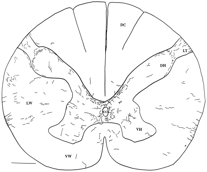
Fig. 10.
Human cervical spinal cord. Camera lucida drawing was made of hypocretin-immunoreactive axons in two adjacent 40 μm sections of a C4–C5 cross-section of a human cord. DH, Dorsal horn; VH, ventral horn; DC, dorsal columns; cc, central canal; LW, lateral white matter; VW, ventral white matter;LT, Lissauer’s tract. Scale bar, 1 mm.

