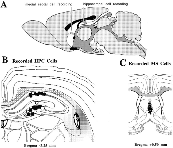
Fig. 1.
A, A diagrammatic representation of the recording arrangement. A tungsten microelectrode was fixed in the region of the stratum moleculare of the dentate on the right side of the brain to record hippocampal field activity (data not shown). Glass microelectrodes carried in independent micromanipulators were simultaneously lowered into the medial septum/vertical limb of the diagonal band of Broca and the hippocampal formation on the left side of the brain, respectively, for the isolation and recording of single cells. B, A diagrammatic reconstruction of cells recorded in the hippocampal formation and classified as theta-related according to the system of Colom and Bland (1987). Seven cells in the CA1 cell body layer (solid circles) and seven cells in the dentate cell body layer (solid circles), for a total of 14 cells, were classified as theta-ON cells (see Results for subclassifications). One cell in the upper blade of the dentate cell body layer was classified as nonrelated (solid square). One cell in the CA1 cell layer (open circle), one cell in the stratum lacunosum region (open circle), and one cell in the upper blade of the dentate cell layer (open circle), for a total of three cells, were classified as theta-OFF cells (see Results for subclassifications).C, A diagrammatic reconstruction of the 18 cells recorded in the medial septal nuclei. All cells were classified as theta-ON (solid circles) and were recorded in a depth range (from the dural surface) of 4.7–5.7 mm. Histology was reconstructed using Swanson’s (1992) atlas.

