
Fig. 5.
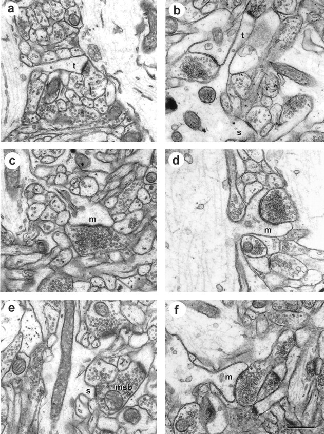
Synapses in area CA1 from perfusion-fixed hippocampus (left) and a hippocampal slice maintainedin vitro for 13 hr before fixation (right). These particular examples were selected to illustrate longitudinally sectioned spines, dendrite origins, and presynaptic boutons. a, Perfusion-fixed thin spine (t). b, In vitrothin (t) and stubby (s) spines. c, Perfusion-fixed mushroom spine.d, In vitro mushroom spine.e, Perfusion-fixed stubby spine on an MSB.f, Another mushroom spine from an in vitro slice. Scale bar: a–f, 0.5 μm.