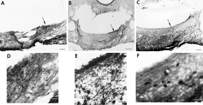
Fig. 6.
Photomicrographs of coronal brain sections showing the transplant containing the SCN in animal B28-Q66 (behavior shown in Fig. 8A). A, The graft lies caudal to the lesion site. There is a plexus of NP staining (arrow) indicating the presence of the donor SCN.B, In a section 50 μm from A, VIP fibers (arrow) overlap the region of the NP fiber plexus; the graft borders are indicated by a dashed line. C, Section adjacent to Bshowing CaBP cells at the same level as the NP and VIP fibers.D–F, Higher magnifications of the areas marked by arrows in A–C, respectively. Scale bars: A–C, 200 μm;D–F, 20 μm.

