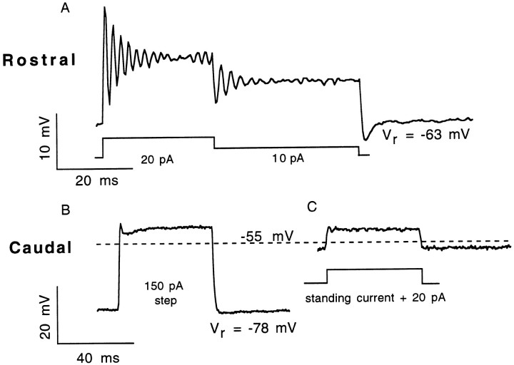
Fig. 2.
Variations of the voltage response of isolated AP hair cells. Current-clamp recordings of the voltage response to a depolarizing current pulse for representative rostral and caudal hair cells. A, Rostral hair cells always exhibited oscillatory responses, and many exhibited membrane potential oscillations at rest. Here, a rostral hair cell is first depolarized by a 20 pA constant current, which is later reduced to a 10 pA current before being turned off. Oscillations occurred at both the onset and transition of the current pulse because the steady-state (direct current) potential was above the threshold forICa. Both resonant frequency andQe were higher at the onset of the 20 pA pulse than at its offset (230 Hz; Qe of 13; steady-state voltage of −52 mV; vs 181 Hz;Qe of 8, Vss of −55 mV). B, Caudal cells typically respond to depolarizing current pulses with a peak that rapidly decays to a plateau, sometimes resembling an overdamped oscillatory response. In this caudal cell, a 150 pA stimulus applied at the zero-current potential of −78 mV of the cell depolarized the membrane potential to a steady-state potential of −53 mV (the potential at whichQe of electrical resonances would typically be highest for oscillatory rostral hair cells). C, The same caudal hair cell with its membrane potential artificially maintained at approximately −55 mV before a 20 pA depolarizing stimulus. Medial hair cell voltage responses could be characterized as one of these two categories (Table 2). Rostral hair cell, 9 pF, 3 MΩ uncompensated series resistance (usr).; caudal hair cell, 4 pF, 1.8 MΩ usr.

