
Fig. 8.
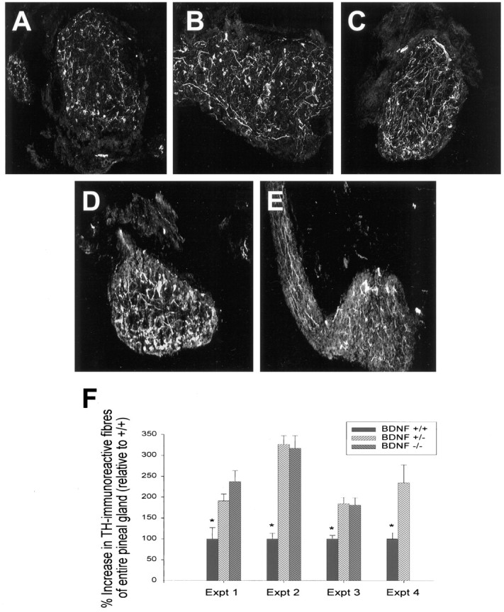
The pineal gland is hyperinnervated with sympathetic fibers in BDNF+/− and BDNF−/− mice at P13. A–C, Immunocytochemical analysis of tyrosine hydroxylase, a specific marker for sympathetic axons, in sections of the pineal gland from (A) BDNF+/+, (B) BDNF+/−, and (C) BDNF−/− littermates. Note that the density of TH-positive fibers is increased in both the BDNF+/− and −/− sections relative to the section from the control littermate. D, E, Immunocytochemical analysis of TH (D) and p75NTR (E) in sections from the same P13 BDNF+/− pineal gland. Note that although the p75NTR-immunoreactivity is somewhat fainter, the pattern of immunoreactivity is similar to that seen with anti-TH . F, Quantitative analysis of the relative amount of pineal gland area covered by TH-immunoreactive fibers in BDNF+/+, +/−, and −/−animals, obtained using sections similar to those shown inA–C. For details of the analysis, see Results and Materials and Methods. Each experiment represents the results obtained from the pineal glands of one set of littermates of different genotypes. Note that in all four experiments, the amount of TH-positive innervation in the BDNF+/+ pineal gland was significantly lower than that seen in either the BDNF+/− or BDNF−/− pineal glands (*p < 0.05). A–E, Magnification, 160×.