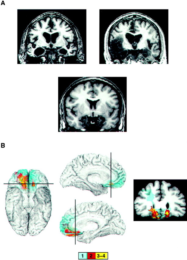
Fig. 1.

Neuroanatomical findings in the two groups of brain-damaged patients. A, Bilateral amygdala lesions. Coronal sections through the amygdala from the three patients in our Registry show complete bilateral destruction of the amygdala. The lesions from the two remaining amygdala patients have been shown in previous publications (Lee et al., 1988a,b, 1995). B, Bilateral VMF lesions. Shown are mesial and inferior views of the overlap of lesions from four VMF patients. The lesions from individual subjects were transferred onto a reference brain by using the MAP-3 technique (Frank et al., 1997). The coronal section shows an area of the ventromedial prefrontal cortex where maximum overlap occurs. The position of the cut is indicated on the brain on theleft. The color bar below shows the color code corresponding to the number of overlapping lesions. The lesion of the fifth VMF patient is not part of the MAP-3 image because, as explained in the text, this patient suffered from a frontal lobe cyst at age 2. The lack of a clear structural lesion at macroscopic level precludes the transfer into MAP-3.
