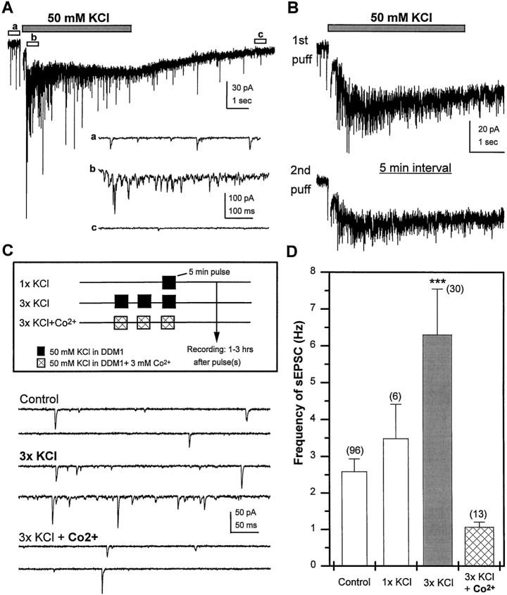
Fig. 11.
Brief depolarization of differentiated wild-type neurons induces calcium-dependent persistent increase in sEPSC frequency. A, A 5 sec, focal application of 50 mm KCl, pressure-ejected from a micropipette ∼20 μm from the cell body, induces a rapid and transient increase in sEPSC frequency riding on a sustained inward current, followed by a decline to below baseline levels within 10 sec. Higher-resolution images of sEPSCs recorded before (a), during (b), and after (c) the KCl puff are indicated below.B, In a single neuron, similar responses are observed to two puffs of KCl when separated by a 5 min rest period in normal external solution. C, Three different paradigms used for global depolarization of cultures at 3–6 DIV by exposure to 50 mm KCl in DDM1. Recordings obtained 1–3 hr after stimulus illustrate an increase in sEPSC frequency after repetitive KCl stimulation that is blocked if stimulation is conducted in the presence of Co2+. D, Mean sEPSC frequency, assayed 1–3 hr after treatment, is plotted for each of the depolarization paradigms. The slightly higher mean sEPSC frequency after 1x KCl was not significantly different from control. However, a significant increase in sEPSC frequency was observed in cultures treated with high 3x KCl when compared with control (ANOVA; ***p < 0.0001, Fisher PLSD). This increase was blocked when Co2+ was present during the repeated exposure to high potassium. Bars indicate SEM, and the number of neurons is indicated in parentheses.

