Fig. 3.
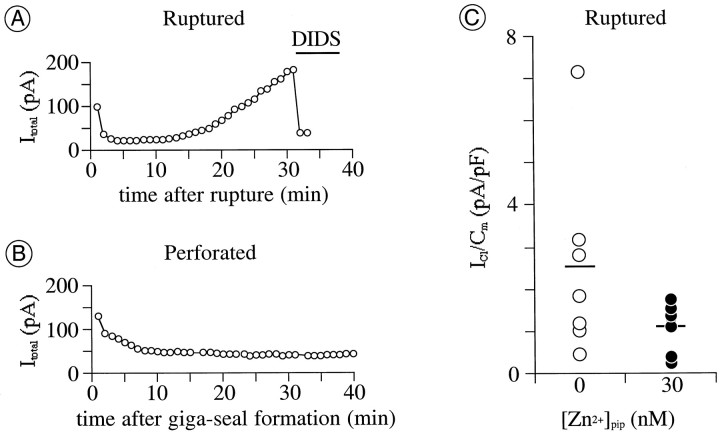
Exogenously supplemented intracellular Zn2+ impedes augmentation ofICl by intracellular perfusion.A, B, Amplitude of total current atEtest of +40 mV plotted against recording time. Recordings in A and B were in ruptured- and perforated-patch mode, respectively. Pipette solutions in both recordings contained no added zinc ([Zn2+]pip = 0); 1 mmDIDS was microperfused onto the cell during the time indicated by thehorizontal bar in A.Cm, 10 pF in A and 28 pF in B. C,ICl activated by depolarization to +40 mV after 12–15 min of intracellular perfusion with Zn2+-free versus Zn2+-containing pipette solutions. Each circle plots the increase in amplitude of ICl over this time for a different cell, normalized by the Cm of that cell. Horizontal bars plot the mean of values for each pipette solution [mean ± SD; 2.6 ± 2.3;n = 7 cells for [Zn2+]pip = 0 nm(open circles); 1.1 ± 0.6; n = 6 for [Zn2+]pip = 30 nm (filled circles)].

