
Fig. 3.
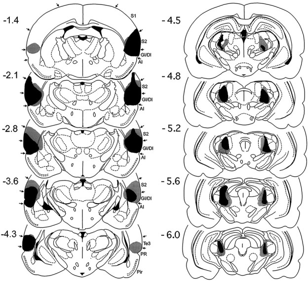
Histological reconstructions of the smallest (black) and largest (gray) combined lesions of posterior parietal insular cortex and posterior intralaminar nuclei in Experiment 2 on coronal plates from the atlas ofPaxinos and Watson (1986). The numbers to the leftindicate rostrocaudal levels relative to bregma.