
Fig. 3.
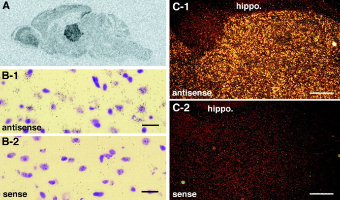
Citron is abundantly expressed in the thalamus.A, Film autoradiography of a parasagittal section of an adult mouse brain examined by in situ hybridization using an antisense riboprobe of Citron mRNA. B-1,B-2, Light-field views of individual thalamic neurons expressing Citron mRNA; most thalamic neurons were positive and associated with numerous silver grains. Scale bars, 20 μm.C-1, C-2, Dark-field view of Citron mRNA expression in the thalamus; note the sharp contrast between the high expression in the thalamus compared with the hippocampal formation (hippo.). Scale bars, 80 μm. In B andC, adjacent sections hybridized using antisense and sense probes are shown.