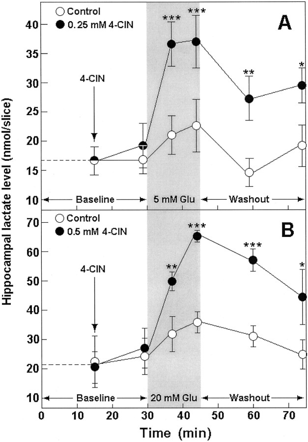
Fig. 1.
Tissue content of lactate before (Baseline) and during (shaded area) exposure to Glu and during Glu washout (Washout) in control (open symbols) and 4-CIN-treated (filled symbols) rat hippocampal slices perfused with either 4 (A) or 10 mm(B) glucose–aCSF. *p < 0.03; **p = 0.01; ***p < 0.004, significantly different from control.

