
Fig. 1.
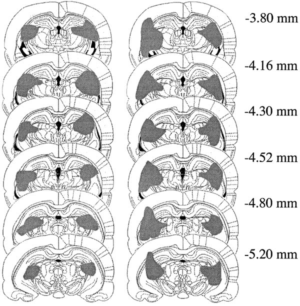
Largest (right) and smallest (left) electrolytic lesions centered on the intergeniculate leaflet in six coronal sections at the levels indicated in millimeters posterior to bregma (from Paxinos and Watson, 1998).
Official websites use .gov
A
.gov website belongs to an official
government organization in the United States.
Secure .gov websites use HTTPS
A lock (
) or https:// means you've safely
connected to the .gov website. Share sensitive
information only on official, secure websites.
Largest (right) and smallest (left) electrolytic lesions centered on the intergeniculate leaflet in six coronal sections at the levels indicated in millimeters posterior to bregma (from Paxinos and Watson, 1998).