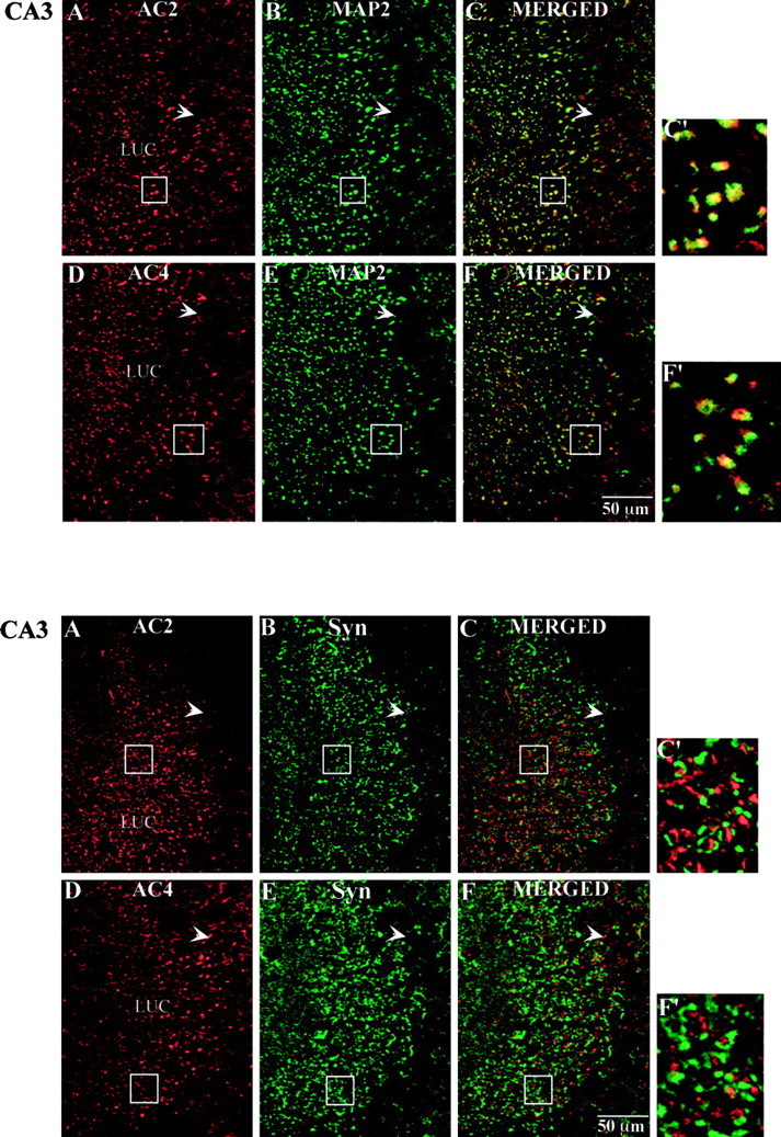
Fig. 10.

Top. Immunohistochemical localization of AC2 and AC4 with MAP2 in the CA3 region of the mouse hippocampus. Mouse brain sections (40 μm) were processed for immunohistochemistry as described in Materials and Methods. A, AC2 labeling (red). B, MAP2 labeling of the same image shown in A (green).C, Merged image of A and B(yellow indicates coincident localization of AC2 and MAP2). D, AC4 labeling (red).E, MAP2 labeling of the same image shown inD (green). F, Merged image of D and E(yellow as described in C).C′, F′, Higher magnification images of the boxed areas in C andF, respectively. Scale bar, 50 μm. Boxed areas in A and B correspond to that inC. Boxed areas in D and Ecorrespond to that in F.
