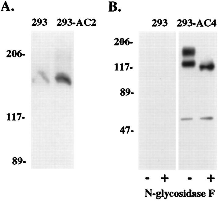
Fig. 3.
Western blot detection of AC2 and AC4 in HEK 293 cell membranes. A, Whole-cell lysate from HEK 293 cells expressing the pCEP4 vector alone (293) or AC2-pCEP4 (293-AC2) was subjected to native PAGE conditions and Western blotted as described in Materials and Methods. Blots were developed using enhanced chemiluminescence and scanned on a Hewlett-Packard Scan Jet II CX scanner. B, Membranes from HEK 293 cells were prepared and treated with or without N-glycosidase F as described in Materials and Methods. Twenty micrograms of membranes from 293-pCEP4 cells (293;lanes 1 and 2) and 293-AC4 cells (293-AC4; lanes 3 and4) were separated by SDS-PAGE on 7.5% acrylamide gels. Protein was transferred to nitrocellulose and incubated with anti-AC4 antibody. Molecular weight markers equal 206, 117, 89, and 47 kDa. Note the conversion of the glycosylated form(s) to the lower molecular weight nonglycosylated form after treatment with N-glycosidase F. A minor band at ∼60 kDa is only seen in AC4-expressing cells and is therefore probably a proteolytic fragment of AC4 that is recognized by the AC4 antibody.

