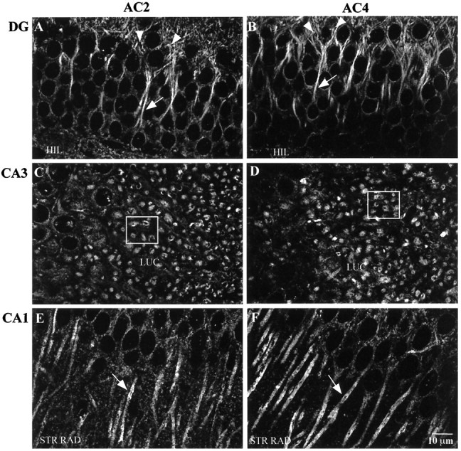
Fig. 5.

Immunohistochemical detection of AC2 and AC4 in the mouse hippocampal formation. Mouse brain sections (40 μm) were processed for immunohistochemistry as described in Materials and Methods. A, AC2 labeling in the dentate gyrus (DG). B, AC4 labeling in dentate gyrus. Note the concentration of AC2 and AC4 label in the proximal dendrites (arrows) and into the dendritic field in the molecular layer (arrowheads). C, AC2 labeling in area CA3. D, AC4 labeling in area CA3. Note the concentration of labeling in the CA3 neuron dendrites in the stratum lucidum (the dendrites are viewed in the coronal plane in these tissue sections). The boxed areas in C andD show specific examples of the described labeling pattern.E, AC2 labeling in area CA1. F, AC4 labeling in area CA1. Note the accumulation of label in the CA1 cell bodies and along the membrane of the CA1 dendrites (arrows) in the stratum radiatum. HIL, Hilus; LUC, stratum lucidum; STR RAD, stratum radiatum. Scale bar, 10 μm.
