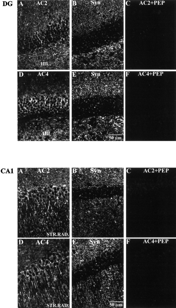
Fig. 8.

Top. Immunohistochemical localization of AC2 and AC4 relative to synaptophysin in the mouse dentate gyrus. Mouse brain sections (40 μm) were processed for immunohistochemistry as described in Materials and Methods. A, AC2 labeling.B, Synaptophysin (Syn) labeling of the same image shown in A. Note that the labeling pattern for AC2 is not coincident with that for synaptophysin.C, Peptide adsorption of AC2 eliminating AC2 labeling in the dentate gyrus. D, AC4 labeling. E, Synaptophysin labeling of the same image shown in D. Note that the labeling pattern for AC4 is not coincident with that for synaptophysin. F, Peptide adsorption of AC4 eliminating AC4 labeling in the dentate gyrus. Scale bar, 50 μm.
