Fig. 7.
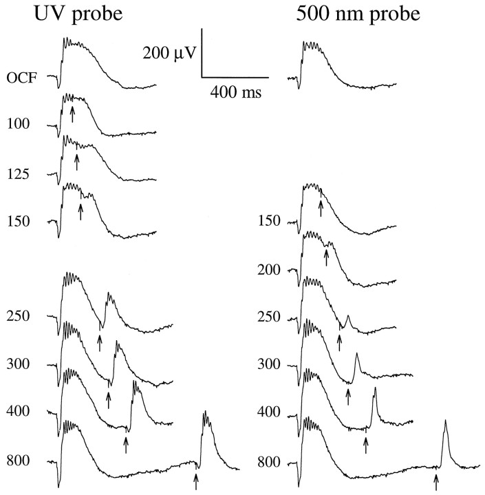
Interaction of signals originating in the UV- and M-cone pigments. Recovery of the cone b-wave after a bright orange (λ > 530 nm; OG-530 colored glass filter) conditioning flash (OCF) estimated to isomerize 1% of the M-cone pigment. Numbers near the traces are the interflash intervals in milliseconds; the trace labeled OCF is the response to the conditioning flash alone (no probe flash).Right-hand traces show responses to a λnom= 500 nm probe flash producing 71,000 photons μm−2 at the cornea. Left-hand traces show responses to a broadband UV probe flash (330–390 nm; Schott UG1 glass) producing ∼43,000 “equivalent” 350 nm photons μm−2 at the cornea; the equivalency is computed with respect to the UV pigment template shown in Figure 6 (Eq.3). The probe flashes were the most intense the apparatus could generate (Fig. 5A, abscissa). (To eliminate most of the rod signal but keep the cones maximally sensitive, the entire experiment was performed in the presence of a steady 520 nm background producing ϕ = 750 photoisomerizations/sec per rod. To insure absence of any UV light in the orange conditioning flash, the intensity of the flash generated by the same flash unit with a combination of Schott glass filters (UG1 + OG-530 + BG39) was measured with a photodiode; the BG39 filter was added to block infrared light. The photodiode failed to record any signal. This indicates that the OCF produced <200 photons μm−2 at the cornea in the UV region of the spectrum.)

