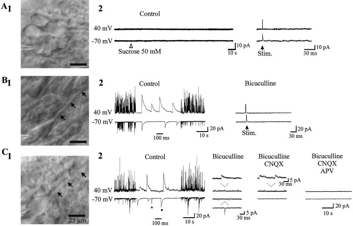
Fig. 5.
Visual identification of silent and active neurons. A1, Silent cell (Cm, 10 pF) with round shape soma and no apical dendrite. A2, Bath application of sucrose (50 mm) or stimulation of the stratum radiatum (100 V, 30 μsec duration) failed to evoke any synaptic response both at −70 and 40 mV. B1, GABA-active neuron (Cm, 27 pF) from the same slice has a bigger soma and an apical dendrite (arrows).B2, The spontaneous activity is completely blocked by bicuculline (10 μm). In the presence of the antagonist, stimulation of the stratum radiatum (100 V, 30 μsec duration) failed to evoke a PSC at −70 and 40 mV (mean of 10 traces evoked at 0.05 Hz). C1, GABA plus Glu-active cell from another slice (Cm, 40 pF). C2, Note that AMPA receptor-mediated PSCs (asterisk) can easily be distinguished from GABA PSCs (filled circle) at −70 mV by the different decay time constant (τ = 1.33 msec, τ = 19.76 msec, respectively).

