
Fig. 1.
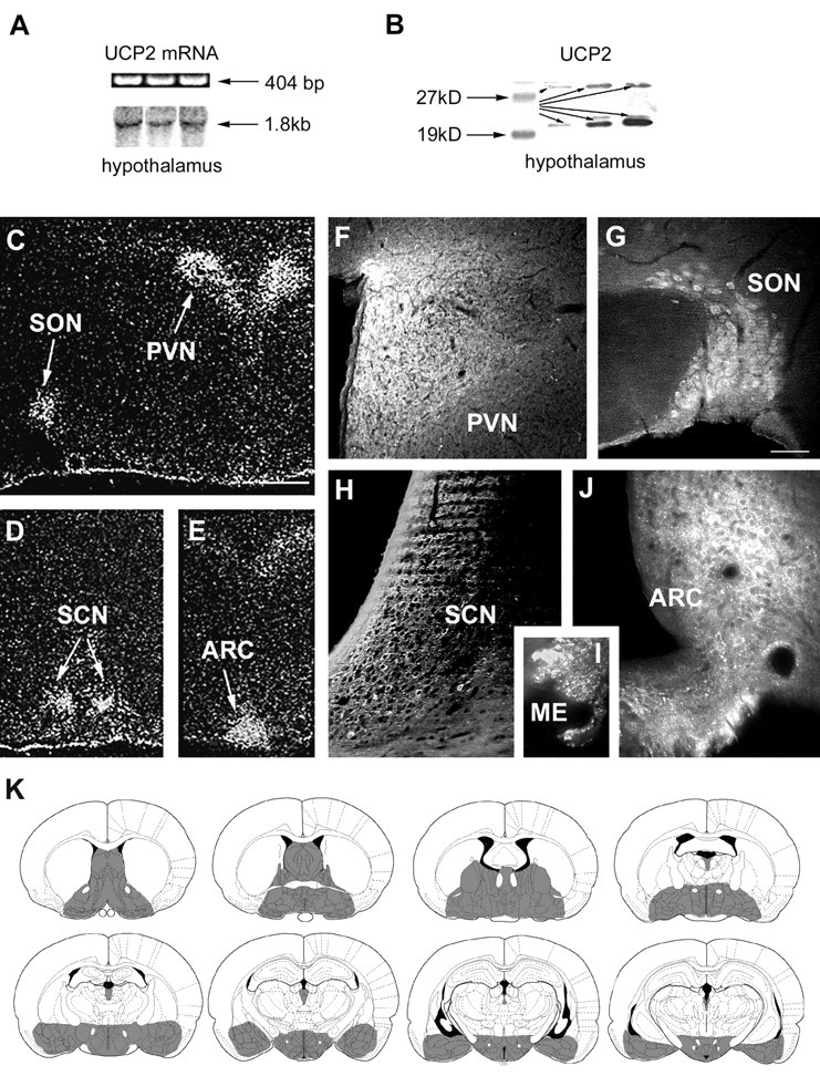
UCP2 mRNA and peptide expression in the brain.A, RT-PCR (top row) and Northern blot analyses of rat hypothalamic tissue demonstrates the expression of UCP2 mRNA. B, Western blot analysis of rat hypothalamic tissue revealed the expression of UCP2 peptide. Two major bands (arrows) were visible using the affinity-purified antisera: a band at 33 kDa that corresponds to the predicted molecular weight of UCP2 and another strong band at 23 kDa was also detected.C–E,In situ hybridization of UCP2 mRNA shows the concentration of labeled cells in four nuclei of the hypothalamus: the paraventricular (PVN), supraoptic (SON), suprachiasmatic (SCN), and arcuate nuclei (ARC).F–I, Although immunolabeling for UCP2 in the rat hypothalamus resulted in perikarya labeling of cells in the nuclei that contained UCP2 mRNA (SON, PVN, SCN, ARC), labeled cellular processes were abundant throughout the hypothalamus. For example, the median eminence (ME) that contains axonal fibers en route to the portal capillaries and the posterior lobe of the pituitary abundantly expressed UCP2. K, Schematic illustration (based on the rat brain atlas of Paxinos and Watson, 1997) of UCP2 (gray shadedareas) in the forebrain and diencephalon based on the in situhybridization and immunocytochemistry studies.