
Fig. 2.
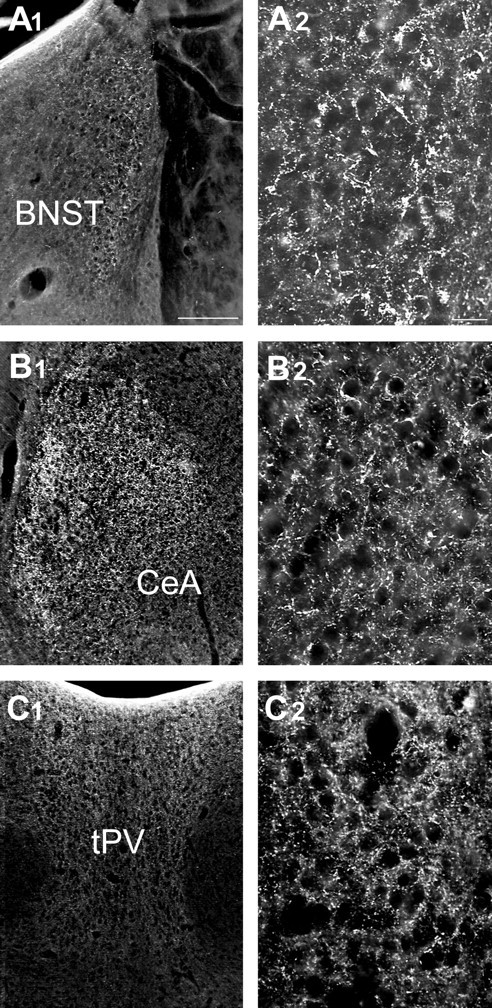
UCP2 in extrahypothalamic limbic sites.A1–C2, Light micrographs reveal the expression of UCP2 immunoreactivity in extrahypothalamic limbic sites. These areas included the bed nucleus of the stria terminalis (BNST; A1,A2), the central nucleus of the amygdala (CeA; B1,B2), and the thalamic paraventricular nucleus (tPV; C1,C2). The higher power magnifications (A2,B2, C2) demonstrate the robust expression of UCP2 in neuronal processes in each of these areas. Scale bars: A1, 100 μm; A2, 25 μm.