
Fig. 3.
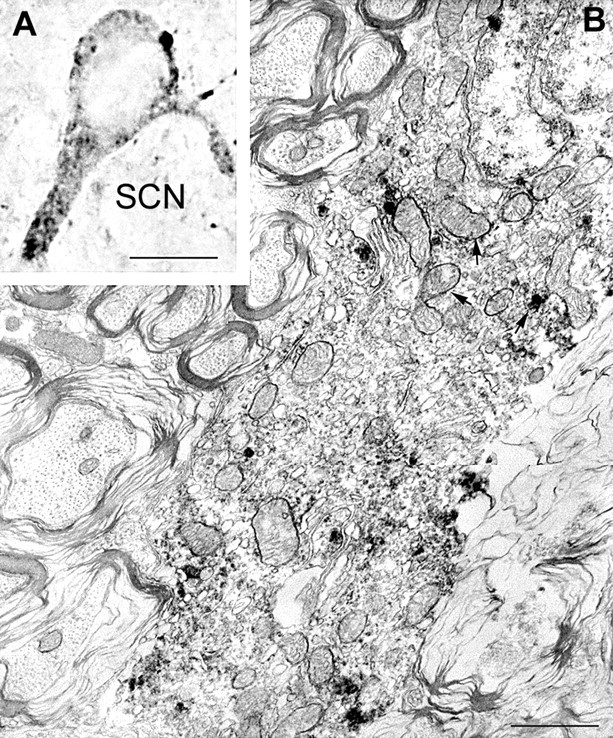
UCP2 in perikaryal mitochondria. Light (A) and electron (B) micrographs demonstrate UCP2 immunolabeling of perikaryal mitochondria (arrows) in the rat SCN. Immunoperoxidase is also associated with cytosol in close apposition to labeled mitochondria. Note the infolded nucleus of this SCN neuron. Scale bars:A, 10 μm; B, 1 μm.