
Fig. 7.
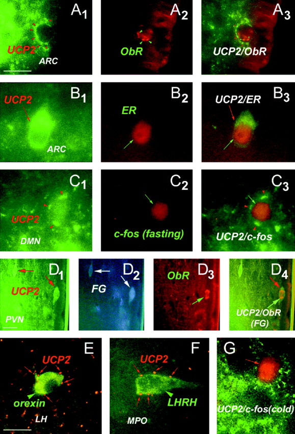
Relationship between UCP2 and neuronal circuits involved in endocrine and metabolic regulation.A1–A3, Green fluorescent UCP2-immunoreactive axon terminals (A1, red arrowheads) in close proximity to an arcuate nucleus neuron expressing immunoreactivity for receptor of the peripheral metabolic hormone leptin (ObR;A2). The image ofA3 was taken using filters sensitive for both red and green fluorescence.B1–B3 , Green fluorescent, UCP2-immunoreactive perikaryonin the arcuate nucleus containing labeling for the peripheral sex hormone estradiol (ER; red fluorescence).C1–C3 , Green fluorescent UCP2-immunoreactive axon terminals (red arrowheads) in close proximity to a perikaryon of the dorsomedial hypothalamic nucleus (DMN) that was activated by fasting (red fluorescent, nuclear c-fos labeling). Fasting-induced c-fos expression was also detected in the lateral hypothalamus-perifornical region, arcuate nucleus, medial preoptic area, central amygdaloid nucleus, and the paraventricular thalamic nucleus.D1–D4 , Green fluorescent UCP2-producing parvicellular neurons of the paraventricular nucleus (PVN;D1, arrows) are neuroendocrine as revealed by retrograde tracing from the periphery using fluorogold (D2 , FG; FG labeling vas visualized using transillumination with ultraviolet light). The UCP2-expressing neuroendocrine cell located to the right(D3, D4,green arrows) also contains leptin receptors (ObR; D3 , red fluorescence). E, Red fluorescent, UCP2-containing axon terminals (red arrows) in close proximity to a neuronal perikaryon (green arrowhead) in the lateral hypothalamus (LH) that expresses the appetite-inducing peptide orexin. F, Red fluorescent, UCP2-containing axon terminals (red arrows) in close proximity to a hypophyseotropic neuron that produce luteinizing hormone-releasing hormone (LHRH) in the medial preoptic region (MPO). G, Green fluorescent UCP2-immunoreactive axon terminals (red arrowheads) in close proximity to a perikaryon of MPO that was activated by cold exposure (red fluorescent, nuclear c-fos labeling). Other cold-activated neurons were distributed in the paraventricular nucleus, medial preoptic area, bed nucleus of the stria terminalis, central amygdaloid nucleus, and the paraventricular thalamic nucleus.