
Fig. 1.
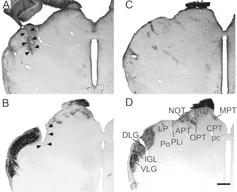
A, GFAP-IR identifying the scar (arrows) of a representative knife-cut thalamus between the dorsomedial dorsal lateral geniculate nucleus and the lateral posterior nucleus. B, CT-β-IR identifying visual projections in the lateral geniculate region, pretectum, and tectum of a representative hamster that received a knife cut (arrows) medial to the dorsal lateral geniculate nucleus. C, GFAP-IR in a control brain. The IGL is indicated by fairly dense GFAP-IR, as are numerous blood vessels.D, Normal visual projections identified with CT-β-IR in a control animal. In both B and D, animals received bilateral intraocular injections of the tracer. Scale bar, 470 μm. APT, Anterior pretectal nucleus;CPT, commissural pretectal nucleus; DLG,dorsal lateral geniculate; IGL, intergeniculate leaflet;LP, lateral posterior nucleus; MPT,medial pretectal nucleus; NOT, nucleus of the optic tract; OPT, olivary pretectal nucleus;pc, posterior commissure; PLi, posterior limitans nucleus; Po, posterior thalamic nucleus;PPT, posterior pretectal nucleus; VLG,ventral lateral geniculate.