Fig. 6.
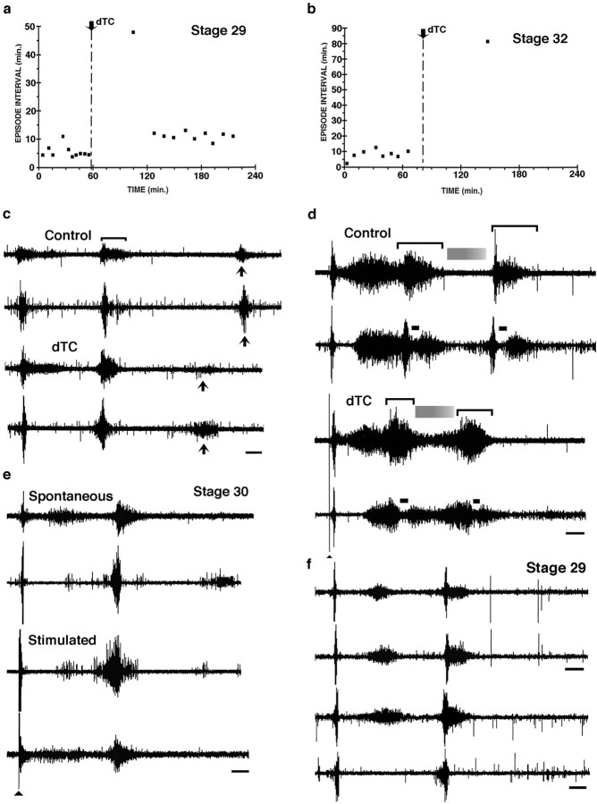
Recording from muscle nerves in isolated spinal cord preparations treated acutely (a–d) and chronically (e, f) with dTC.a, Scatter plot showing the intervals between spontaneous bursting episodes in a st. 29 embryo that, under control conditions, burst every 5.6 ± 2.1 min (mean ± SD;n = 10). The addition of dTC transiently blocked bursting, which then resumed at a lower frequency; the time between episodes was 14.4 ± 1.29 min (mean ± SD;n = 9). b, Scatter plot showing the interval between spontaneous bursting episodes in a st. 32 embryo; under control conditions an episode occurred every 8.2 ± 3.03 min (mean ± SD; n = 8), whereas after the addition of dTC only a single episode occurred after 82 min and none occurred with an additional 50 min. c, In the same embryo as in a, the entire bursting episode before dTC is shown (top pair oftraces; femorotibialis and sartorius, respectively); a single burst is indicated by a bracket. Thebottom pair of traces shows the burst structure of spontaneous episodes after the recovery of bursting in the continued presence of 5 × 10⁻⁶mdTC. Note the changes in burst structure: increased amplitude of the first two bursts and the decrease in the third burst and its time of appearance (compare solid arrows).d, The structure and length of the episode (the same embryo as in b) are altered from control (top pair of traces; femorotibialis and sartorius, respectively) after the addition of 5 × 10⁻⁶mdTC (bottom pair of traces; femorotibialis and sartorius, respectively; see Results for more details). Brackets indicate the two bursts occurring in each episode; shaded grayrectanglesindicate a quiescent period between bursts, and the solid bar indicates an inhibitory period that occurs in sartorius at the onset of each burst. Solid arrowhead indicates a stimulus artifact for lower two traces only.e, In a chronically treated dTC embryo in the continuous presence of 5 × 10⁻⁶mdTC, both spontaneous and stimulated bursting episodes occur. Femorotibialis (top) and sartorius (bottom) are shown in each pair of traces.f, Neurogram recordings from embryos treated with endo-N plus dTC. The top pair oftraces is from a st. 29 embryo chronically treated withdTC and recorded in the continued presence of 5 × 10⁻⁶mdTC. The burst structure of the femorotibialis from the limb injected with endo-N at the onset of the chronic dTC treatment (lower trace) is essentially the same as that recorded from the femorotibialis in the noninjected hindlimb. The lower pair of traces is from an embryo that was chronically injected with endo-N alone. Both traces from the endo-N-injected limb (top trace, femorotibialis;bottom trace, sartorius) when 5 × 10⁻⁶mdTC was acutely added to the bath show that the burst structure is similar to the embryo chronically treated with dTC (top pair of traces) and that this differed from the burst structure before dTC treatment (as shown in Fig.5a). Thus acute dTC treatments produce the same effects as chronic dTC treatments, and this is not affected by the presence or absence of endo-N. Calibration bars:c–f, 1 sec.

