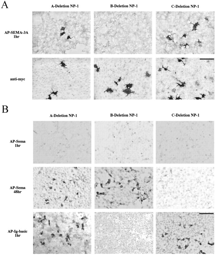
Fig. 2.
Mapping SEMA-3A binding sites to neuropilin-1 domains. A, HEK293T cells were transiently transfected with A-, B-, or C- deletion neuropilin-1 and probed with ∼1.5 nm AP-SEMA-3A (top) or an anti-myc antibody (bottom). After 1 hr development in NBT/BCIP, AP-SEMA-3A was visualized bound to cells expressing A-deletion and C-deletion neuropilin-1 but not to those expressing B-deletion neuropilin-1. Staining live cells with anti-myc demonstrates that all constructs are expressed on the cell surface. B, The same neuropilin-1 deletion constructs were probed with ∼3 nm AP-Sema (top two rows) or 1.5 nm AP-Ig-basic (bottom). After 2 d development, AP-Sema was visualized bound to A- and B-deletion neuropilin-1 but not to C-deletion neuropilin-1. After 1 hr development, AP-Ig-basic was visualized bound to A- and C-deletion neuropilin-1 but not B-deletion neuropilin-1. Scale bars: A, 62.5 μm;B, 100 μm.

