
Fig. 1.
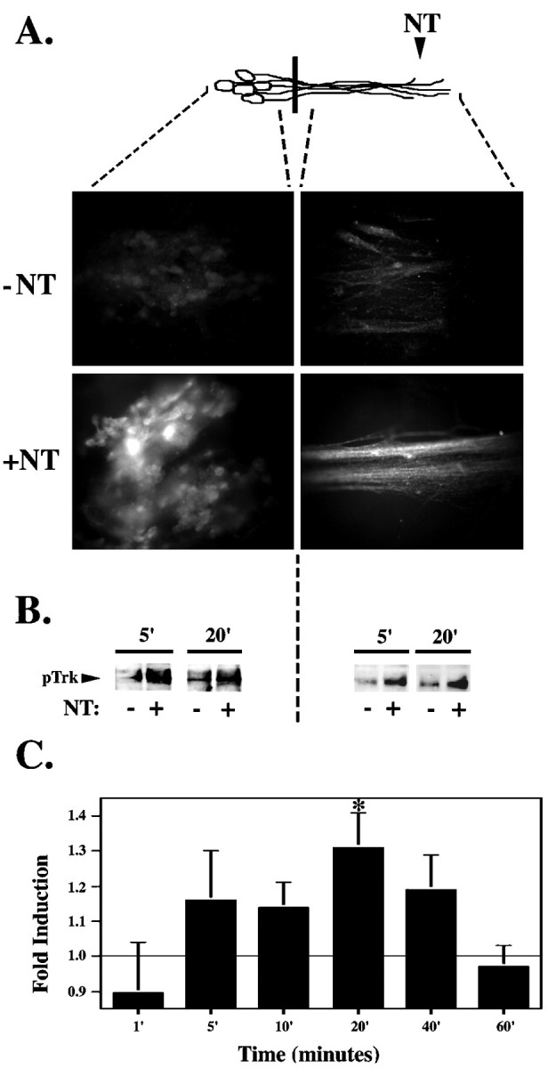
Trk is rapidly activated in response to neurotrophin stimulation. A, Selective neurotrophin stimulation of DRG neurites results in pTrk immunostaining in neurites and cell bodies of DRG neurons. After a 20 min neurotrophin (+NT) or vehicle control (−NT) (0.1 ng/ml BSA) stimulation at the neurites, DRG neurons were fixed in 4% paraformaldehyde and immunostained using the anti-pTrk antibody in conjunction with a Cy3-conjugated secondary antibody. B, Trk phosphorylation is detected in the cell body as early as 5 min after selective neurotrophin (+NT) or vehicle control (−NT) (0.1 ng/ml BSA) stimulation at the neurites. DRG neuron lysates from the neurites and from the cell bodies were pooled from nine compartmented cultures, separated on 10% SDS-PAGE gel, transferred to Immobilon-P membrane, and immunoblotted with anti-pTrk. C, Time course of Trk phosphorylation at the cell body in response to neurite stimulation. Intensity of the pTrk band was quantitated with the Molecular Dynamics Storm 860 imaging system using Image Quant (version 2.0; Molecular Dynamics) and normalized for protein loading. The time course of pTrk induction (calculated as the ratio between pTrk in neurotrophin- and control-stimulated cultures) was calculated from several experiments (n = 3–10 for each time point; *p < 0.05). Diagram (top) is a schematic representation of neurons; black bar indicates presence of the Teflon divider. Area of DRG neurons analyzed is indicated by dashed lines.