Fig. 5.
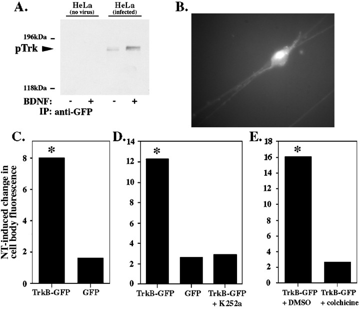
pTrk localized to the neurites is transported to the cell body. A, Uninfected HeLa cells (lanes 1, 2) or HeLa cells expressing TrkB-GFP (lanes 3, 4) were treated with PBS control (−) or 100 ng/ml BDNF (+) for 5 min. Cell lysates were immunoprecipitated with anti-GFP, followed by immunoblotting with anti-pTrk. B, Three days after infection of DRG neurons, expression of TrkB-GFP is visible in the cell bodies and the neurites as fluorescence. C–E, DRG cell bodies of neurons expressing TrkB-GFP grown in compartmented cultures were bleached until the fluorescence was reduced by 90% (C) or left unbleached (D, E). Fluorescence emitted by the cell bodies was measured after addition of 100 ng/ml BDNF and 100 ng/ml NGF or vehicle control to the neurites. Cell body fluorescence at 10 min after neurite stimulation was calculated as a percent of cell body fluorescence at time 0. Neurotrophin-induced change in cell body fluorescence equals mean change in cell body fluorescence in neurotrophin-stimulated cultures minus mean change in cell body fluorescence in control-stimulated cultures. The experiment was repeated using DRG neurons expressing GFP alone (C,D). The kinase inhibitor K252a was applied to neurites 30 min before neurotrophin stimulation of DRG neurons expressing TrkB-GFP (D). To test whether the TrkB-GFP is transported from the neurites to the cell bodies, we repeated the experiment in the presence of colchicine or a vehicle control (DMSO). Colchicine (125 μm) or DMSO was applied to neurites 60 min before neurotrophin stimulation (E). The percent change in fluorescence over the course of 10 min was compared between the neurotrophin- and control-stimulated cells using a two tailed t test assuming unequal variance (n >15 for each point; *p < 0.05).

