
Fig. 3.
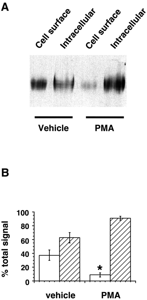
PKC activation results in DAT redistribution from the cell surface to an intracellular pool. A, Steady-state biotinylation of DAT-PC12 cells after 30 min treatment with either vehicle or 1.0 μm PMA. Biotinylated (cell surface) and nonbiotinylated (intracellular) proteins were separated with streptavidin beads and analyzed by immunoblot with a DAT-specific antibody as described in Materials and Methods. A representative immunoblot is shown. B, Quantitation of DAT-PC12 cell biotinylation. Immunoblots of biotinylated (cell surface; open bars) and nonbiotinylated (intracellular; hatched bars) proteins were scanned and quantitated using ImageQuant software. *p < 0.05, significant difference compared with vehicle-treated cells; unpaired Student’st test; n = 4.