
Fig. 1.
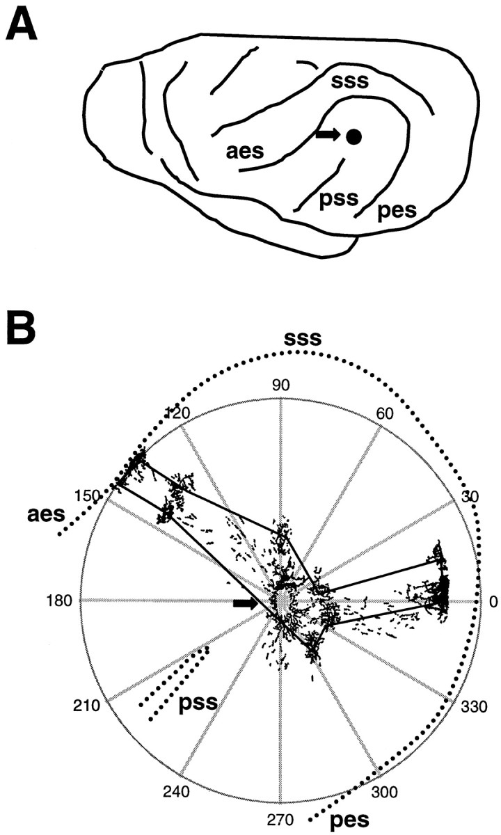
A, Drawing of the left side of a ferret brain, with the location of the sulci surrounding AI noted. Thearrow shows the approximate location of the tracer injections. aes, Anterior ectosylvian sulcus;pes, posterior ectosylvian sulcus; sss,suprasylvian sulcus; pss, pseudosylvian sulcus.B, Illustration of methods used for making polar plots of bouton distribution. The arrow shows the tracer injection site, and the camera lucida drawing of the resulting label is superimposed on the polar plot. With the injection site as the origin, the polar coordinates of the center of each bouton cluster were recorded. Lines were drawn through the center of each bouton cluster to arrive at the plots shown in Figures 6 and 7. Thedashed lines indicate the sulci surrounding AI and show how the plots were oriented.