Fig. 5.
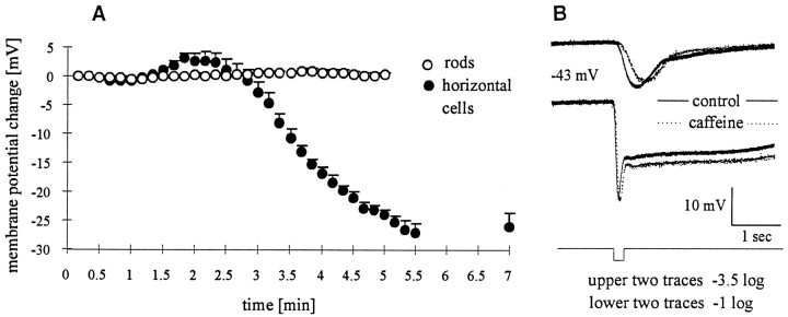
Caffeine hyperpolarizes horizontal cells but not rods. Intracellular recordings from rods and HCs in the eyecup preparation are shown. A, Caffeine (10 mm) was added at t = 0 min. No effect on the membrane potential of rods was discerned (open circles), whereas horizontal cells (filled circles) had a two-phased response—transient depolarization followed by a peak hyperpolarization of 26 ± 2.5 mV (n = 7). The effects of caffeine were reversible. In five of six cells recorded for >20 min after caffeine washout, the membrane potential returned to within 3 mV of prestimulus values. B, Light responses of a rod to 200 msec, 567 nm flash (top, −3.5 log;bottom, −1.0 log quanta) before and during caffeine exposure are shown. In caffeine, slightly slower rise times were observed for dim but not for stronger flashes. No significant effect of caffeine was observed on either the transient hyperpolarization of the rod or the rod “tail” for brighter flashes. Plotted membrane potentials were normalized to the resting potential before caffeine.

