Fig. 1.
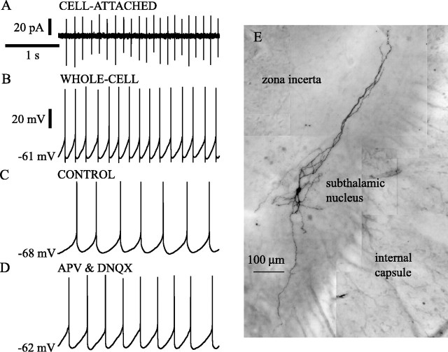
Subthalamic neurons were spontaneously activein vitro. A, B, Example of patch-clamp recordings from a subthalamic neuron that was recorded in the cell-attached configuration (A) before establishing the whole-cell configuration (B). Note the slow (5–7 Hz), rhythmic generation of action currents(A) and action potentials (B).C, D, Intrinsic membrane properties underlie the spontaneous discharge of subthalamic neurons in vitro. A spontaneously active subthalamic neuron (C) continued to fire rhythmically when excitatory amino acid neurotransmission was blocked by the application of the selective NMDA and AMPA receptor antagonists APV and DNQX, respectively (D). E, Composite image based on multiple light micrographs of a subthalamic neuron that was visualized using histochemical procedures (see Materials and Methods) after being recorded in the whole-cell configuration. Time calibration inA applies to A–D. Current calibration inA applies to that figure. Voltage calibration inB applies to B–D. The membrane potential value printed at the left of each traces in this and all subsequent figures refers to the first point in the trace.

