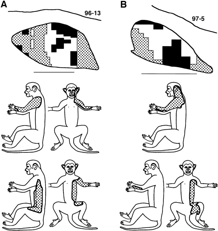
Fig. 12.
Postinjury CN representations of tactile inputs from the forelimb–trunk–face in two maps (A, B).Top, Camera lucida tracings of two CNs. Dorsal =up; medial = right. Scale bar, 1 mm.Stippling indicates CN areas that contained recording sites with forelimb, trunk, and face receptive fields. These areas are located lateral and medial to the dorsal hand representation (white) and adjacent to areas with recording sites that were not responsive to tactile stimulation of the skin (black). Middle, Composite of the receptive field areas of all recording sites with forelimb–trunk–face fields that were located in the area lateral to the hand representation for the above CN. In each case, note that this skin included the forelimb, shoulder, neck, and face. Bottom, Composite of the receptive field areas of all recording sites with forelimb–trunk fields that were located medial to the hand representation for the above CN. In each case, note that this skin included the forelimb, trunk, and proximal hindlimb.

