
Fig. 6.
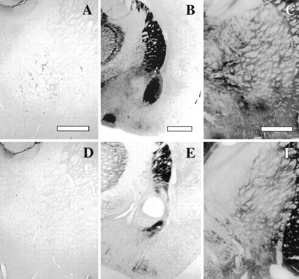
Confirmation of the location and extent of the lesions used in Experiment 2. A, D, p75 immunostaining of NBM magnocellular neurons in a control and immunolesioned brain, respectively. Scale bar, 600 μm. B, E, AChE histochemistry at the level of the basolateral amygdala in a control and NMDA-induced intra-amygdala lesion, respectively. Scale bar, 1 mm.C, F, AChE histochemistry in the basal forebrain in a control and NMDA-induced intrabasal lesion, respectively. Scale bar, 600 μm.