
Fig. 1.
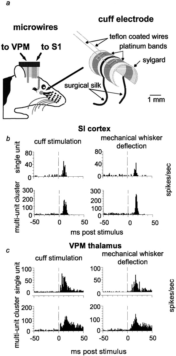
Nerve cuff electrode design and comparison of sensory responses in VPM and SI resulting from nerve cuff stimulation and manual whisker deflection. a, Schematic diagram of nerve cuff electrode and microwire placement. b, c, Peristimulus time histograms of responses to nerve cuff stimulation and manual whisker deflection from the same recording session in anesthetized rats. Examples of a single-unit and a multiunit recording are shown for VPM (c) and SI (b). Note that b andc were collected from two separate animals.Vertical dashed lines indicate the time of stimulus presentation (0 msec).