Fig. 1.
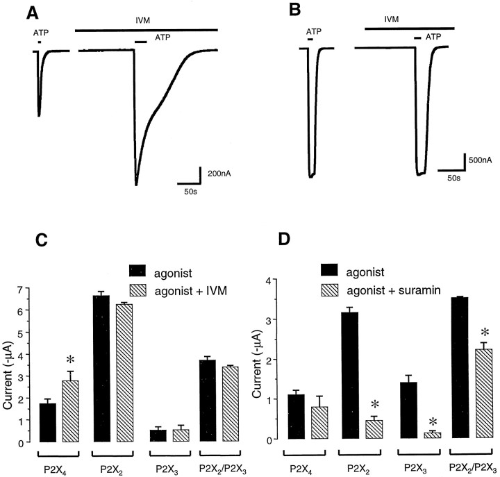
IVM potentiates P2X4 but not P2X2 or P2X3 channel currents.A, Representative recordings of currents mediated by P2X4 channels expressed in oocytes. Left trace, 100 μm ATP-evoked current before and after (right trace) addition of IVM (10 μm); IVM potentiates the amplitude as well as duration of the ATP-evoked current. B, Representative recordings for P2X2 channels expressed in oocytes. Left trace, 100 μm ATP-evoked current before and after (right trace) addition of IVM (10 μm); IVM causes no change in either the holding current or ATP-evoked current in P2X2-expressing cells. C, Summary of data from a number of cells (n > 5 for each) showing that IVM (10 μm) potentiates ATP-evoked currents at P2X4 channels but not at P2X2, P2X3, and P2X2/P2X3channels. D, Summary of data from a number of cells (n > 5 for each) showing that suramin (30 μm) can block ATP-evoked currents at P2X2, P2X3, and P2X2/P2X3 channels but not at P2X4 channels. Subsequent figures show a more pronounced potentiation of P2X4 channels at lower IVM and/or ATP concentrations. The data are derived from measurements of peak ATP-evoked currents.

